Selection Criteria In Dental Radiography
The Office of Communication, Education, and Radiation Programs (OCER) in conjunction with the American Dental Association has updated the 1987 FDA publication The Selection of Patients for X-Ray Examinations: Dental Radiographic Examinations. The recommendations in this updated document provide the dentist with a strategy for ordering x-ray examinations for patients seeking treatment while at the same time minimizing radiation exposure. The revised recommendations update information on newer technologies, update the scientific literature and terminology, describe the use of radiographs when assessing various types of patients and discuss the use of protective aprons when taking radiographs.These revised recommendations were developed to serve as an adjunct to the dentist's professional judgment of how to best use diagnostic imaging for each patient. Radiographs can help the dental practitioner evaluate and definitively diagnose many oral diseases and conditions. However, the dentist must weigh the benefits of taking dental radiographs against the risk of exposing a patient to x-rays, the effects of which accumulate from multiple sources over time.
The dentist, knowing the patient's health history and vulnerability to oral disease, is in the best position to make this judgment in the interest of each patient. For this reason, these recommendations are intended to serve as a resource for the practitioner and are not intended to be a standard of care, requirements or regulations.The documents are available from the links listed below and from the web site.
Also available at: Revised 2012AMERICAN DENTAL ASSOCIATIONCouncil on Dental Benefit ProgramsCouncil on Scientific Affairs U.S. DEPARTMENT OF HEALTH AND HUMAN SERVICESPublic Health ServiceFood and Drug Administration CONTENTS.BackgroundThe dental profession is committed to delivering the highest quality of care to each of its individual patients and applying advancements in technology and science to continually improve the oral health status of the U.S. These guidelines were developed to serve as an adjunct to the dentist’s professional judgment of how to best use diagnostic imaging for each patient. Radiographs can help the dental practitioner evaluate and definitively diagnose many oral diseases and conditions. However, the dentist must weigh the benefits of taking dental radiographs against the risk of exposing a patient to x-rays, the effects of which accumulate from multiple sources over time. The dentist, knowing the patient’s health history and vulnerability to oral disease, is in the best position to make this judgment in the interest of each patient.
Selection Criteria For Dental Radiography 3rd Edition
For this reason, the guidelines are intended to serve as a resource for the practitioner and are not intended as standards of care, requirements or regulations.The guidelines are not substitutes for clinical examinations and health histories. The dentist is advised to conduct a clinical examination, consider the patient’s signs, symptoms and oral and medical histories, as well as consider the patient’s vulnerability to environmental factors that may affect oral health. This diagnostic and evaluative information may determine the type of imaging to be used or the frequency of its use. Dentists should only order radiographs when they expect that the additional diagnostic information will affect patient care. Based on this premise, the guidelines can be used by the dentist to optimize patient care, minimize radiation exposure and responsibly allocate health care resources.
1IntroductionThe guidelines titled, 'The Selection of Patients for X-Ray Examination' were first developed in 1987 by a panel of dental experts convened by the Center for Devices and Radiological Health of the U.S. Food and Drug Administration (FDA). The development of the guidelines at that time was spurred by concern about the U.S.
Population’s total exposure to radiation from all sources. Thus, the guidelines were developed to promote the appropriate use of x-rays. In 2002, the American Dental Association, recognizing that dental technology and science continually advance, recommended to the FDA that the guidelines be reviewed for possible updating.
The FDA welcomed organized dentistry’s interest in maintaining the guidelines, and so the American Dental Association, in collaboration with a number of dental specialty organizations and the FDA, published updated guidelines in 2004. This report updates the 2004 guidelines and includes recommendations for limiting exposure to radiation.Patient Selection CriteriaRadiographs and other imaging modalities are used to diagnose and monitor oral diseases, as well as to monitor dentofacial development and the progress or prognosis of therapy.
Radiographic examinations can be performed using digital imaging or conventional film. The available evidence suggests that either is a suitable diagnostic method. 2-4 Digital imaging may offer reduced radiation exposure and the advantage of image analysis that may enhance sensitivity and reduce error introduced by subjective analysis.


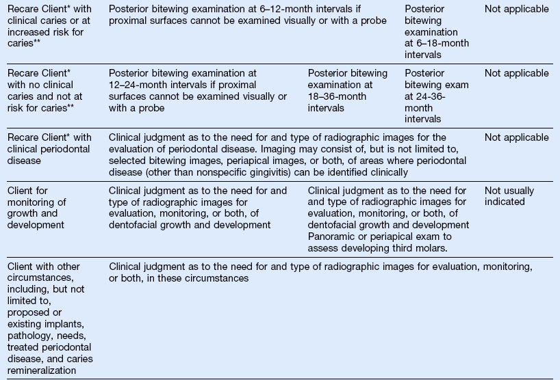
5A study of 490 patients found that basing selection criteria on clinical evaluations for asymptomatic patients, combined with selected periapical radiographs for symptomatic patients, can result in a 43 percent reduction in the number of radiographs taken without a clinically consequential increase in the rate of undiagnosed disease. Table 1 TYPE OF ENCOUNTERPATIENT AGE AND DENTAL DEVELOPMENTAL STAGEChild with Primary Dentition (prior to eruption of first permanent tooth)Child with Transitional Dentition (after eruption of first permanent tooth)Adolescent with Permanent Dentition(prior to eruption of third molars)Adult, Dentate or Partially EdentulousAdult, EdentulousNew Patient. being evaluated for oral diseasesIndividualized radiographic exam consisting of selected periapical/occlusal views and/or posterior bitewings if proximal surfaces cannot be visualized or probed. Patients without evidence of disease and with open proximal contacts may not require a radiographic exam at this time.Individualized radiographic exam consisting of posterior bitewings with panoramic exam or posterior bitewings and selected periapical images.Individualized radiographic exam consisting of posterior bitewings with panoramic exam or posterior bitewings and selected periapical images. Table 2 EquipmentFrequencyMethodX-ray MachineOn installationAt regular intervals as recommended by state regulationsWhenever there are any changes in installation workload or operating conditionsInspection by qualified expert (as specified by government regulations and manufacturers recommendations).Film ProcessorOn installationDailyMethod 1: Sensitometry and Densitometry A sensitometer is used to expose a film, followed by standard processing of the film.